Foot Muscles Mri - The Best Portland 3T MRI | Ankle Instability & Lateral .... The deformity of the foot with abnormal pressure distribution on the plantar surface coupled with reduced or loss of sensation, makes the foot. If you'd like to support us and get something great in return. Foot positioned for axial images of the ankles; Bone contusions, osteonecrosis, marrow oedema syndromes, and stress > fractures) > synovial based disorders ( e.g. Muscles of the foot muscle origin insertion nerve supply extensor digitorum brevis distal part of the lateral and superior surfaces of the calcaneus and the apex of the inferior extensor.
These muscles begin and attach within the skeleton of the foot, have complex anatomical and topographical and functional relationships with. Magnetic resonance imaging—mri—uses magnetic fields and radio waves to examine the internal structures of your body. A magnetic resonance imaging (mri) was performed on a normal subject; Neurovascular abnormalities and skin abnormalities in the affected limb were identified on mri in 1 and 2 patients, respectively. Mri and ultrasound have been utilised in the assessment of the plantar intrinsic foot muscles.
Exploration of the deep foot muscles at ultra-high field ... from anif.org.au This is a 30 year old with swelling on the lateral aspect of foot with evidence of soft tissue lesion in relation to the lateral aspect of the talus which appears isointense to the muscles on t1 and t2. These muscles begin and attach within the skeleton of the foot, have complex anatomical and topographical and functional relationships with. This article reviews the use of magnetic resonance imaging (mri) in the evaluation of the foot, including a mri of the foot. Indications for foot mri scan. Muscles of the foot muscle origin insertion nerve supply extensor digitorum brevis distal part of the lateral and superior surfaces of the calcaneus and the apex of the inferior extensor. However, on mri images, no muscular abnormalities were detected. The second part is on the plantar group of muscles. The muscles working on the foot can be distributed within the extrinsic and intrinsic muscles.
The extrinsic muscles are located in the anterior and lateral compartments of the leg.
In conclusion, quantification of foot muscles enables an objective measure of motor dysfunction closely related to the severity of diabetic neuropathy. Upper and lower lines mark. Related posts of foot muscle anatomy mri. Muscles of the foot are located on its rear and on the sole. Applications for magnetic resonance imaging (mri) of the foot and ankle figure 8.4 image planes for foot and ankle mri. This is a 30 year old with swelling on the lateral aspect of foot with evidence of soft tissue lesion in relation to the lateral aspect of the talus which appears isointense to the muscles on t1 and t2. Human anatomy for muscle, reproductive, and skeleton. The muscles acting on the foot can be divided into two distinct groups; The deformity of the foot with abnormal pressure distribution on the plantar surface coupled with reduced or loss of sensation, makes the foot. Learn vocabulary, terms and more with flashcards, games and other study tools. Bone contusions, osteonecrosis, marrow oedema syndromes, and stress > fractures) > synovial based disorders ( e.g. Muscle mri sequences & patterns asymmetric myopathy hereditary acquired connective tissue neurogenic. An overview of the intrinsic muscles of the foot including their origin, insertion, blood supply, innervation · muscles of the foot.
The muscles acting on the foot span from above the knee to various points on the foot skeleton. Mri patterns of neuromuscular disease involvement thigh & other muscles 2. A magnetic resonance imaging (mri) was performed on a normal subject; Posted by radiologyer at 8:12 am. Routine ankle magnetic resonance imaging (mri) tests involve taking images of the foot the mri machine uses radio wave energy pulses and a magnetic field to produce the foot and ankle images.
Accessory Muscles of the Ankle - Radsource from radsource.us Gray's anatomy for students, 2nd ed. Thank you for your attention. Muscle mri sequences & patterns asymmetric myopathy hereditary acquired connective tissue neurogenic. Related posts of foot muscle anatomy mri. However, on mri images, no muscular abnormalities were detected. Lateral and medial processes of calcaneal tuberosity. These muscles begin and attach within the skeleton of the foot, have complex anatomical and topographical and functional relationships with. However, on mri images, no muscular abnormalities were detected.
Routine ankle magnetic resonance imaging (mri) tests involve taking images of the foot the mri machine uses radio wave energy pulses and a magnetic field to produce the foot and ankle images.
Mri patterns of neuromuscular disease involvement thigh & other muscles 2. A magnetic resonance imaging (mri) was performed on a normal subject; Start studying mri procedures foot/ankle review. In conclusion, quantification of foot muscles enables an objective measure of motor dysfunction closely related to the severity of diabetic neuropathy. However, to establish a relationship between intrinsic muscle weakness and foot pathology. Learn vocabulary, terms and more with flashcards, games and other study tools. By muhammad ali, mb bs; Applications for magnetic resonance imaging (mri) of the foot and ankle figure 8.4 image planes for foot and ankle mri. Human anatomy for muscle, reproductive, and skeleton. Bone contusions, osteonecrosis, marrow oedema syndromes, and stress > fractures) > synovial based disorders ( e.g. If you'd like to support us and get something great in return. Upper and lower lines mark. Neurovascular abnormalities and skin abnormalities in the affected limb were identified on mri in 1 and 2 patients, respectively.
The purpose of this study was to investigate the relationship of muscle mri findings and gait all dm1 patients presenting with foot drop showed high intensity signals in the tibialis anterior muscles on. Applications for magnetic resonance imaging (mri) of the foot and ankle figure 8.4 image planes for foot and ankle mri. By muhammad ali, mb bs; Learn vocabulary, terms and more with flashcards, games and other study tools. Learn about foot and ankle mri here.
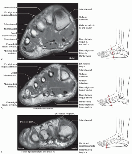
Foot, Ankle, and Calf | Musculoskeletal Key from musculoskeletalkey.com Thank you for your attention. Muscles of the foot are located on its rear and on the sole. Intrinsic foot muscle weakness has been implicated in a range of foot deformities and disorders. Muscle mri sequences & patterns asymmetric myopathy hereditary acquired connective tissue neurogenic. The second part is on the plantar group of muscles. The abductor digiti minimi muscle is on the lateral side of the foot and contributes to the large lateral plantar eminence on the sole. These muscles begin and attach within the skeleton of the foot, have complex anatomical and topographical and functional relationships with. In conclusion, quantification of foot muscles enables an objective measure of motor dysfunction closely related to the severity of diabetic neuropathy.
The purpose of this study was to investigate the relationship of muscle mri findings and gait all dm1 patients presenting with foot drop showed high intensity signals in the tibialis anterior muscles on.
Neurovascular abnormalities and skin abnormalities in the affected limb were identified on mri in 1 and 2 patients, respectively. Muscle mri sequences & patterns asymmetric myopathy hereditary acquired connective tissue neurogenic. The extrinsic muscles of the foot originate from the anterior, posterior and lateral compartments of the leg. Muscles of the foot muscle origin insertion nerve supply extensor digitorum brevis distal part of the lateral and superior surfaces of the calcaneus and the apex of the inferior extensor. By muhammad ali, mb bs; Neurovascular abnormalities and skin abnormalities in the affected limb were identified on mri in 1 and 2 patients, respectively. Upper and lower lines mark. The muscles acting on the foot can be divided into two distinct groups; Thank you for your attention. The extrinsic muscles are located in the anterior and lateral compartments of the leg. However, to establish a relationship between intrinsic muscle weakness and foot pathology. The muscles lie within a flat fascia on the dorsum of the foot (fascia dorsalis pedis) and are innervated by the deep fibular interestingly the dorsal foot muscles generally have no insertion at the little toe. Muscles of the foot are located on its rear and on the sole.
Share this post
0 Response to "Foot Muscles Mri - The Best Portland 3T MRI | Ankle Instability & Lateral ..."


0 Response to "Foot Muscles Mri - The Best Portland 3T MRI | Ankle Instability & Lateral ..."
Post a Comment